Cancer continues to be one of the world’s most complex health challenges, where early detection, precise diagnosis, and accurate surgical planning can dramatically influence patient outcomes. As medical imaging technologies advance, Computer Vision—especially segmentation—has become a critical enabler of the next generation of cancer detection and diagnostic tools.
At Visual Grab Computer Vision IT Services, our expertise lies in building powerful AI models that strengthen the intelligence layer of cancer detection software. We focus exclusively on model development—ensuring that companies building cancer-tech platforms have the highest-quality deep learning foundation to succeed.
1. Understanding the Landscape: Major Types of Cancer
Cancer is not a single disease; it encompasses many forms depending on the types of cells involved. Accurate segmentation and detection techniques vary across cancer types due to differing imaging characteristics.
1.1 Carcinomas
These begin in epithelial tissues and constitute the majority of cancer diagnoses:
Breast cancer
Lung cancer
Colorectal cancer
Prostate cancer
Skin cancer (melanoma & non-melanoma)
Most imaging modalities—mammography, CT, MRI, dermoscopy—require advanced segmentation to locate lesions accurately.
1.2 Sarcomas
Rare and diverse tumors arising from connective tissues such as:
Bone (osteosarcoma)
Fat (liposarcoma)
Muscle (rhabdomyosarcoma)
Segmenting these tumors is more complex due to irregular shapes and heterogeneous textures.
1.3 Leukemias
Cancers of blood-forming tissues, often analyzed using:
Digital blood smear microscopy
AI-driven segmentation helps isolate white blood cells and detect malignant transformations.
1.4 Lymphomas
Affecting the lymphatic system, these cancers rely heavily on:
CT
PET
MRI
Segmentation helps identify enlarged lymph nodes and differentiate malignant from benign swellings.
1.5 Central Nervous System (CNS) Tumors
Includes:
Gliomas
Astrocytomas
Meningiomas
Brain tumor segmentation is one of the most challenging tasks due to:Diffuse boundaries
Edema regions
Tumor heterogeneity
1.6 Pediatric Cancers
Cancers like neuroblastoma, Wilms’ tumor, and retinoblastoma require highly sensitive segmentation models as early identification significantly improves survival.
2. How Segmentation Supports Cancer Detection & Surgical Precision
2.1 Developing High-Accuracy Early Detection Systems
Segmentation isolates abnormal tissue from:
MRI
CT
PET
X-Ray
Ultrasound
Histopathology slides
These segmented regions help algorithms:
Detect cancer early
Reduce false negatives
Prioritize cases for radiologists
Enable automated screening workflows
2.2 Assisting Radiologists With Clearer Interpretation
Segmentation algorithms offer:
Clear boundaries of suspicious lesions
Tumor volume measurements
Progression tracking
Consistency across radiologists and scans
This improves diagnosis speed and accuracy.
2.3 Powering Surgical Planning & Navigation
Precision segmentation is essential in:
Brain tumor surgeries
Breast-conserving procedures
Liver resections
Lung nodule removal
Surgeons rely on models that:
Generate 3D reconstructions
Highlight vital structures to avoid
Estimate margins of resection
Reduce risk of recurrence
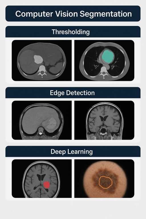
3. Computer Vision Segmentation Approaches: Techniques & Benefits
Segmentation techniques have evolved dramatically. Below are key approaches relevant to cancer detection.
3.1 Traditional Segmentation Methods
Thresholding & Region-Based Segmentation
Works well for high-contrast images
Extremely fast
Suitable for simple tumor boundaries
Edge Detection Methods
Sobel, Canny, Laplacian
Good for structural delineation
Often used as a preprocessing step
Classical ML Methods
k-Means
Random Forests
Watershed
Graph Cuts
Useful where datasets are small or when interpretability is required.
3.2 Deep Learning–Based Segmentation (The Industry Standard)
U-Net & U-Net Variants
Most widely used for biomedical imaging
Performs exceptionally on small datasets with augmentation
High pixel-level accuracy
Mask R-CNN
Performs detection and segmentation simultaneously
Excellent for histopathology imaging
Handles overlapping tumors
DeepLab v3/v3+
Handles complex boundaries
Multi-scale feature extraction
Transformer-Based Models
Swin UNet
SegFormer
Offer:Powerful global context
Better handling of irregular tumor shapes
3D CNN Architectures
Used for volumetric CT/MRI data where depth information is essential.
Vital for:
Brain tumors
Lung nodules
Liver metastases
3.3 Semi-Supervised & Weakly Supervised Segmentation
Helps when annotated medical datasets are scarce:
Uses unlabeled data efficiently
Reduces annotation cost
Improves generalization
This is crucial in cancer imaging where expert labeling is expensive.
4. Why Segmentation Quality Determines the Success of Cancer Detection Software
Building cancer detection software requires more than classification—it demands high-precision segmentation because:
Tumor shapes are irregular
Small lesions may be life-threatening
Clinical decisions rely on exact boundaries
Volumetric measurements require pixel-perfect accuracy
Surgical plans depend on precise region isolation
A weak segmentation model leads to:
Missed cancers
Wrong staging
Incorrect treatment planning
Reduced trust from clinicians
This is why segmentation is the core intelligence layer of cancer diagnostics.
5. How Visual Grab Helps Companies Build High-Quality Cancer Detection Models
At Visual Grab, we work exclusively on AI model R&D—building, training, refining, and optimizing segmentation and detection models that your product team can integrate into your own clinical workflows.
We do not handle:
Compliance (FDA / CE)
PACS/HIS integration
Deployment or on-site implementation
Our mission is clear:
We build exceptional models. You build the healthcare product.
✔ End-to-End Model Development (Research → Prototype → High-Accuracy Models)
Our capabilities include:
Tumor segmentation
Organ segmentation
Lesion localization
Multi-class segmentation
3D volumetric model development
Histology slide segmentation
Multi-modality fusion models
We engineer datasets, design architectures, and build robust training pipelines.
✔ Advanced Deep Learning Architecture Implementation
We work with:
U-Net family
Mask R-CNN
DeepLab
Swin UNet / SegFormer (Transformers)
3D CNNs and hybrid models
We choose architectures based on:
Imaging modality
Tumor type
Complexity
Availability of labeled data
✔ Full Training Pipeline Setup
We handle:
Data augmentation
Loss function optimization
Class imbalance challenges
Curriculum learning
Ensemble techniques
Hyperparameter tuning
Each training workflow is built to maximize segmentation accuracy and stability.
✔ Model Evaluation, Benchmarking & Reporting
We provide detailed reports with metrics like:
Dice Score
IoU
Precision & Recall
Volumetric error
Boundary error metrics
Each report helps your engineering team validate the model internally and prepare for regulatory processes (handled by your own compliance teams).
**✔ Model Optimization for Real-World Deployment
(Optimization only — deployment done by your engineers)**
We optimize models for:
Speed
Memory
High-resolution images
Stability across scanners and settings
Your engineering team receives integration-ready AI models.
6. Conclusion: Building the AI Core of Tomorrow’s Cancer Detection Systems
The future of cancer detection will rely on advanced segmentation models that understand medical images at unprecedented levels of detail. Companies building cancer detection software need AI engines that are:
Precise
Reliable
Interpretable
Scalable
At Visual Grab Computer Vision IT Services, our role is to build those engines.
We partner with MedTech innovators who want to create world-changing cancer detection solutions—and we power them with the deep learning excellence they need to make it possible.
**Want to build the AI core of your cancer detection product?
Let’s collaborate and accelerate your vision.**